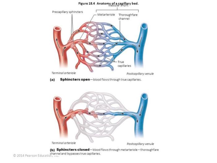
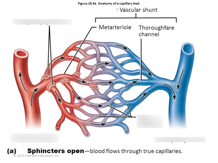
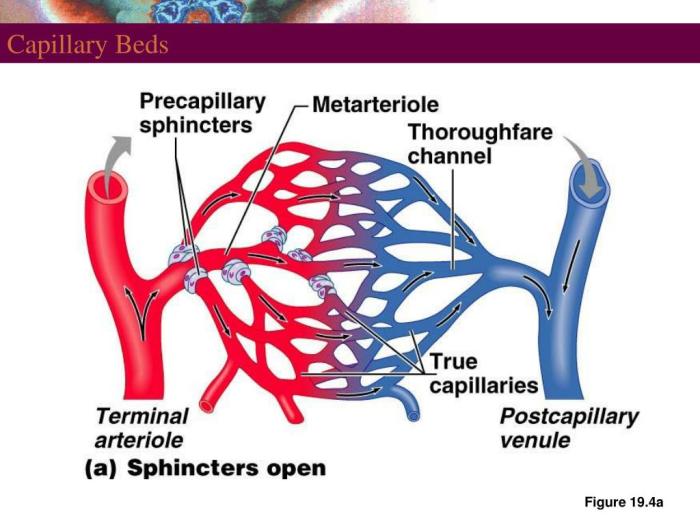
Label the structures of the capillary bed – Labeling the structures of the capillary bed unveils a fascinating realm of microcirculation, providing insights into the intricate network that facilitates vital physiological processes. This comprehensive guide delves into the types, structure, functions, and labeling methods of capillaries, showcasing their significance in capillary biology and advancing our understanding of human physiology.
Capillaries, the smallest blood vessels, play a crucial role in nutrient and oxygen exchange, waste removal, and immune responses. Understanding their structure and function is essential for unraveling the complexities of human biology and developing targeted therapies for various diseases.
Structures of the Capillary Bed: Label The Structures Of The Capillary Bed

Capillaries are the smallest and most numerous blood vessels in the body. They form a network that connects arteries to veins and allow for the exchange of oxygen, nutrients, and waste products between the blood and the surrounding tissues.
Types of Capillaries
There are three main types of capillaries:
- Continuous capillaries: These are the most common type of capillary. They have a continuous layer of endothelial cells that are joined by tight junctions. This prevents the leakage of fluid and solutes from the blood into the surrounding tissues.
- Fenestrated capillaries: These capillaries have small pores in their endothelial cells. This allows for the leakage of fluid and solutes from the blood into the surrounding tissues.
- Sinusoidal capillaries: These capillaries have large pores in their endothelial cells. This allows for the leakage of fluid, solutes, and even blood cells from the blood into the surrounding tissues.
Structure of a Typical Capillary
A typical capillary is a thin-walled tube with a diameter of about 5-10 micrometers. The walls of the capillary are made up of a single layer of endothelial cells. These cells are joined by tight junctions, which prevent the leakage of fluid and solutes from the blood into the surrounding tissues.
The endothelial cells of capillaries are also covered by a basement membrane. This is a thin layer of connective tissue that helps to support the capillary walls.
Functions of the Different Capillary Structures
The different types of capillary structures have different functions. Continuous capillaries are found in tissues where the exchange of fluid and solutes is tightly controlled. Fenestrated capillaries are found in tissues where the exchange of fluid and solutes is less tightly controlled.
Sinusoidal capillaries are found in tissues where the exchange of fluid, solutes, and even blood cells is necessary.
Methods for Labeling Capillary Structures

There are a variety of methods that can be used to label capillary structures. These methods include:
- Immunohistochemistry: This method uses antibodies to label specific proteins in the capillary walls.
- Lectin histochemistry: This method uses lectins to label specific carbohydrates in the capillary walls.
- Electron microscopy: This method can be used to visualize the ultrastructure of capillary walls.
Advantages and Disadvantages of Each Method
Each of these methods has its own advantages and disadvantages. Immunohistochemistry is a relatively simple and inexpensive method that can be used to label a wide variety of proteins. However, it can be difficult to find antibodies that are specific for the proteins of interest.
Lectin histochemistry is a simple and inexpensive method that can be used to label a wide variety of carbohydrates. However, it can be difficult to find lectins that are specific for the carbohydrates of interest.
Electron microscopy is a powerful method that can be used to visualize the ultrastructure of capillary walls. However, it is a relatively expensive and time-consuming method.
Step-by-Step Protocol for Labeling Capillary Structures Using a Specific Method
The following is a step-by-step protocol for labeling capillary structures using immunohistochemistry:
- Collect tissue samples and fix them in a suitable fixative.
- Embed the tissue samples in paraffin or plastic.
- Cut thin sections of the tissue samples.
- Apply the primary antibody to the tissue sections.
- Wash the tissue sections to remove unbound antibody.
- Apply the secondary antibody to the tissue sections.
- Wash the tissue sections to remove unbound antibody.
- Visualize the labeled capillary structures using a microscope.
Applications of Capillary Labeling

Capillary labeling can be used to study a variety of aspects of capillary function. These applications include:
- Studying the distribution of capillaries in different tissues.
- Investigating the changes in capillary structure that occur in response to disease.
- Developing new methods for treating diseases that affect capillaries.
How Capillary Labeling Can Be Used to Study Capillary Function, Label the structures of the capillary bed
Capillary labeling can be used to study a variety of aspects of capillary function. For example, it can be used to measure the rate of fluid and solute exchange between the blood and the surrounding tissues. It can also be used to investigate the role of capillaries in the regulation of blood pressure.
Examples of How Capillary Labeling Has Been Used to Advance Our Understanding of Capillary Biology
Capillary labeling has been used to advance our understanding of capillary biology in a number of ways. For example, it has been used to show that capillaries are not simply passive conduits for the transport of blood. Rather, they play an active role in the regulation of blood flow and the exchange of fluid and solutes between the blood and the surrounding tissues.
FAQs
What are the different types of capillaries?
There are three main types of capillaries: continuous, fenestrated, and sinusoidal. Continuous capillaries are found in most tissues and have a continuous endothelial lining with tight junctions. Fenestrated capillaries have small pores in their endothelial lining, allowing for the passage of small molecules.
Sinusoidal capillaries are found in the liver and spleen and have large, irregular pores that allow for the passage of large molecules and even cells.
What are the functions of capillaries?
Capillaries play a crucial role in the exchange of nutrients, oxygen, and waste products between the blood and tissues. They also help to regulate blood pressure and body temperature.
How are capillary structures labeled?
Capillary structures can be labeled using a variety of methods, including immunohistochemistry, lectin histochemistry, and electron microscopy.
